Retinal Tear Oct : Vitreous Syneresis An Impending Posterior Vitreous Detachment Pvd / Capturing peripheral pathologies can also assist in the differentiation of lesions previously misidentified.
byAdmin-
0
Retinal Tear Oct : Vitreous Syneresis An Impending Posterior Vitreous Detachment Pvd / Capturing peripheral pathologies can also assist in the differentiation of lesions previously misidentified.. Retinal tears when a retinal tear or hole hasn't yet progressed to detachment, your eye surgeon may suggest one of the following procedures to prevent retinal detachment and preserve vision. Ten to 15 percent of patients with acute symptomatic pvd are found to have a retinal tear. A dark shadow blocking part of your peripheral vision. Typically, however, the vitreous separates without any ill effects on the retina. Tears and detachments can be treated with laser surgery or an advanced freezing process.
Sudden onset of eye floaters. They can be classified into tears, such as horseshoe tears and giant retinal tears, or holes, such as operculated holes and atrophic holes. The patient underwent laser retinopexy to barricade the retinal tear. Typically, however, the vitreous separates without any ill effects on the retina. Aging, eye trauma, eye surgery, or being drastically nearsighted may cause retinal tears or detachments.
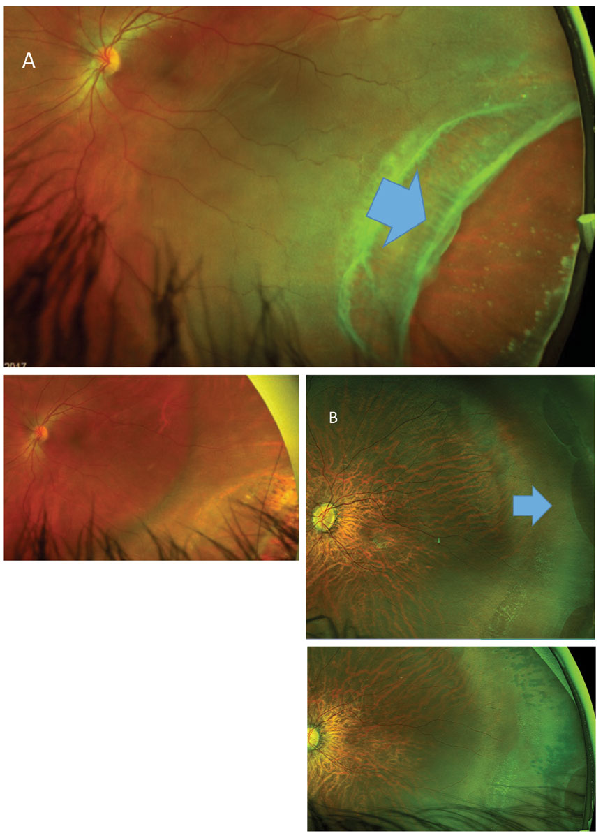
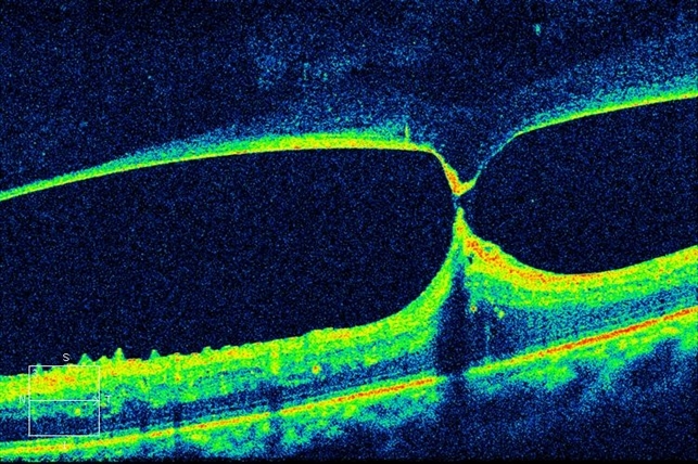
A Field Guide To Retinal Holes And Tears from www.reviewofoptometry.com A retinal tear can lead to fluid and blood collecting in the eye, which can cause the development of several new floaters and loss of vision if the tear leads to a retinal detachment. Atrophic retinal hole (red arrows) noted both on (a) fundus photograph and (b) oct. (b) oct findings after 1 day in an eye with surgery using dispersive ovd (arrows) (dispersive ovd group). The retina plays a vital role in vision. Oct raster scan reveals a subclinical retinal tear with minimal surrounding subretinal fluid in a patient with acute onset vitreous hemorrhage and proliferative dr. Although a tear can usually be repaired successfully, it is considered a serious condition. The areas where the retina detaches lose their blood supply and stop working, causing you to lose vision. They are caused by a hole or tear in the retina that allows fluid to pass through and collect underneath the retina, detaching it from its underlying blood supply.
A retinal tear can allow the liquid part of the vitreous to escape behind the retina and separate the retina from its underlying attachments (and blood supply).
The patient underwent laser retinopexy to barricade the retinal tear. 3,4 vitreous hemorrhage is an important sign. Aging, eye trauma, eye surgery, or being drastically nearsighted may cause retinal tears or detachments. Typically, however, the vitreous separates without any ill effects on the retina. Symptomatic retinal breaks a symptomatic retinal break is defined as a break in the presence of new or increased flashes and/or floaters. A retinal tear can lead to fluid and blood collecting in the eye, which can cause the development of several new floaters and loss of vision if the tear leads to a retinal detachment. If you suspect that it's happened to you, you should consult an optician for a comprehensive eye exam as soon as possible. Posterior vitreous detachment posterior vitreous detachment(pvd) occurs as a natural aging progress but can lead to the risk for severe visual impairment if associated with retinal tears that can then develop into a retinal detachment. The areas where the retina detaches lose their blood supply and stop working, causing you to lose vision. This can have the appearance of someone shaking pepper in your vision. However, occasionally, the flap may be pulled free (avulsed) and be seen as an elongated operculum floating above the break. Retina of the eye functions much more like a film in a camera. Chan ck, meyer ch, gross jg, abraham p, nuthi as, kokame gt et al.
Flap or horseshoe retinal tear a flap (horseshoe) tear results from vitreous traction that pulls a tear of sensory retina that almost always remains attached at the anterior margin of the break. Tears and detachments can be treated with laser surgery or an advanced freezing process. Posterior vitreous detachment posterior vitreous detachment(pvd) occurs as a natural aging progress but can lead to the risk for severe visual impairment if associated with retinal tears that can then develop into a retinal detachment. Chiang a, chang lk, yu f, sarraf d. Retinal tears when a retinal tear or hole hasn't yet progressed to detachment, your eye surgeon may suggest one of the following procedures to prevent retinal detachment and preserve vision.
Tractional Retinal Detachment Retina Image Bank from imagebank.asrs.org Flap or horseshoe retinal tear a flap (horseshoe) tear results from vitreous traction that pulls a tear of sensory retina that almost always remains attached at the anterior margin of the break. Rhegmatogenous detachments are caused by a hole or tear in the retina that allows fluid to pass through and collect underneath the retina, pulling the retina away from underlying tissues. They can be classified into tears, such as horseshoe tears and giant retinal tears, or holes, such as operculated holes and atrophic holes. In addition, if the laser is coded as 67145, and happens today or tomorrow, modifier 57 would also be needed in addition to modifier 24. Oct is heavily used by ophthalmologists to obtain high resolution images of the eye retina. Common retinal tear symptoms include: Chan ck, meyer ch, gross jg, abraham p, nuthi as, kokame gt et al. The retina plays a vital role in vision.
They are caused by a hole or tear in the retina that allows fluid to pass through and collect underneath the retina, detaching it from its underlying blood supply.
This can have the appearance of someone shaking pepper in your vision. Determine macular status in retinal detachment However, occasionally, the flap may be pulled free (avulsed) and be seen as an elongated operculum floating above the break. Chiang a, chang lk, yu f, sarraf d. Sudden onset of eye floaters. A dark shadow blocking part of your peripheral vision. They are caused by a hole or tear in the retina that allows fluid to pass through and collect underneath the retina, detaching it from its underlying blood supply. Chan ck, meyer ch, gross jg, abraham p, nuthi as, kokame gt et al. If any doubt, a retinal oct can demonstrate a detachment easily. Rhegmatogenous (reg ma todge uh nus) retinal detachments are the most common type. Retinal tears can have many causes and can happen at any age. (a) oct findings on day 7 in the control group.the retinal tear is open and curled, and retinal detachment persists; This is known as a rhegmatogenous retinal detachment.
A retinal tear can lead to fluid and blood collecting in the eye, which can cause the development of several new floaters and loss of vision if the tear leads to a retinal detachment. Optical coherence tomography (oct) findings after vitrectomy with application of dispersive ophthalmic viscosurgical device (ovd). Damage to the retina can cause vision loss and even permanent blindness. This is known as a rhegmatogenous retinal detachment. In addition, if the laser is coded as 67145, and happens today or tomorrow, modifier 57 would also be needed in addition to modifier 24.
Retinal Holes And Tears Recognizing Pathology Optos from recognizingpathology.optos.com The retina plays a vital role in vision. However, occasionally, the flap may be pulled free (avulsed) and be seen as an elongated operculum floating above the break. The areas where the retina detaches lose their blood supply and stop working, causing you to lose vision. Oct images can be used to diagnose many retina related eyes diseases. A retinal tear is a small break in this inner lining. Typically, however, the vitreous separates without any ill effects on the retina. A retinal tear (horseshoe) represents vitreoretinal traction and often can lead to retinal detachment, hence the need for prophylactic treatment. (b) oct findings after 1 day in an eye with surgery using dispersive ovd (arrows) (dispersive ovd group).
The patient underwent laser retinopexy to barricade the retinal tear.
3,4 vitreous hemorrhage is an important sign. Atrophic retinal hole (red arrows) noted both on (a) fundus photograph and (b) oct. Chan ck, meyer ch, gross jg, abraham p, nuthi as, kokame gt et al. A retinal tear can occur when the retina pulls away from the outer layers of the eye. Retinal tears can have many causes and can happen at any age. Common retinal tear symptoms include: The ring of pigmentation (blue arrows) is a reactive repair due to separation of neurosensory retina and the retinal pigment epithelium. Rhegmatogenous (reg ma todge uh nus) retinal detachments are the most common type. Aging, eye trauma, eye surgery, or being drastically nearsighted may cause retinal tears or detachments. The areas where the retina detaches lose their blood supply and stop working, causing you to lose vision. Yes, because the care for the retinal tear eye is unrelated to the postoperative care for the slt. Chiang a, chang lk, yu f, sarraf d. However, occasionally, the flap may be pulled free (avulsed) and be seen as an elongated operculum floating above the break.
Chiang a, chang lk, yu f, sarraf d retinal tear. This can have the appearance of someone shaking pepper in your vision.